Sponsored by Hand Therapy New Zealand , the Australian Hand Therapy Association, and Tindeq
Search Results
901 results found with an empty search
- Combined interventions for thumb OA: Are they superior to education alone?
Efficacy of a combination of conservative therapies vs an education comparator on clinical outcomes in thumb base osteoarthritis: A randomized clinical trial. Deveza, L. A., et al. (2021). Level of Evidence : 1b Follow recommendation : 👍 👍 👍 👍 Type of study : Therapeutic Topic : Thumb osteoarthritis - Combined interventions vs self-management and joint protection This is a randomised, single-centre, double-blind, placebo controlled trial assessing the effectiveness of combined interventions vs self-management and ergonomics on pain and function in participants with thumb osteoarthritis (OA). Participants (N = 204) were included if they presented with thumb pain in half of the past month days, pain in 1st cmcj OA of at least 40 out of 100, Functional Index of Hand OA (FIHOA) of at least 6 out of 30, Kellgren-Lawrence grade 2 or higher on x-ray of the 1st cmcj. Participants were excluded if they had had hand surgery or cortisone injections in their hands in the last 6 months. Unfortunately, participants were not excluded if they had previously trialed interventions (e.g. splinting) which were being tested in the study (see full inclusion and exclusion criteria here ). Effectiveness of intervention was assessed through pain (VAS) and function (FIHOA) at baseline, and 6 weeks. Participants and assessors were blinded to treatment allocation. Participants were randomised to either a 6 weeks combined interventions or educational program. The combined intervention program included education, joint protection advice, a neoprene splint (prefabricated neoprene worn for at least 4hrs during the day - see picture below), pain-free hand exercises (e.g. thumb opposition, pinch strengthening, grip strengthening) three times per week, and topical NSAIDs (n = 102). The education only program received education and joint protection advice alone (n = 102). Both groups attended 2 in person sessions (at baseline and at 2 weeks). The results showed that pain improved to clinically significant level in both group without differences between groups at the 6 weeks follow up. Function improved in both groups, however, there was a statistical and potentially clinically relevant difference between groups (favoring the combined interventions) at 6 weeks. Disclaimer: This publication was reviewed and assessed by one reviewer only and it reflects their interpretation. Readers should come to their own conclusions by reading the original article. Clinical Take Home Message : Based on what we know today, combined interventions for thumb OA do not provide greater pain-relief than education and joint protection in people with thumb OA. It is however possible that our clients could get some relevant improvements in hand function in the short term with a combined interventions approach. Considering that a recent large multi-crentred RCT found splinting to have no greater effect than a placebo splint in thumb OA, we may provide our clients with education, exercise, and topical NSAIDs alone. This would allow, to provide our clients with two sessions of hand therapy where we can progress exercises and reiterate key information (the price would be similar to one session of hand therapy + a splint). In addition, we may move away from joint protection programs for hand OA as these have not been shown to be effective in hand OA . Instead we could encourage joint motion for lotion , promote joint movement for amusement , and suggest meditation for elation . If this is not enough and clients want something passive (no exercises) that has been shown to have some effect (compared to placebo), although small, look at supplements for osteoarthritis . Also remember: keep smiling , your clients' pain will decrease! URL : https://doi.org/10.1001/jamainternmed.2020.7101 Available through EBSCO Health Databases for PNZ members. Abstract IMPORTANCE: A combination of conservative treatments is commonly used in clinical practice for thumb base osteoarthritis despite limited evidence for this approach. OBJECTIVE: To determine the efficacy of a 6-week combination of conservative treatments compared with an education comparator. DESIGN, SETTING, AND PARTICIPANTS: Randomized, parallel trial with 1:1 allocation ratio among people aged 40 years and older with symptomatic and radiographic thumb base osteoarthritis in a community setting in Australia. INTERVENTIONS: The intervention group (n = 102) received education on self-management and ergonomic principles, a base-of-thumb splint, hand exercises, and diclofenac sodium, 1%, gel. The comparator group (n = 102) received education on self-management and ergonomic principles alone. Intervention use was at participants' discretion from 6 to 12 weeks. MAIN OUTCOMES AND MEASURES: Hand function (Functional Index for Hand Osteoarthritis; 0-30) and pain (visual analog scale; 0-100 mm) were measured at week 6 (primary time point) and week 12. An α of .027 was used at week 6 to account for co-primary outcomes. RESULTS: Of the 204 participants randomized, 195 (96%) and 194 (95%) completed follow-ups at 6 and 12 weeks, respectively; the mean (SD) age of the population was 65.6 (8.1) years, and 155 (76.0%) were female. At week 6, hand function improved significantly more in the intervention group than the comparator (between-group difference, -1.7 units; 97.3% CI, -2.9 to -0.5; P = .002). This trend was sustained at 12 weeks (-2.4 units; 95% CI, -3.5 to -1.3; P < .001). Pain scores improved similarly at week 6 (between-group difference, -4.2 mm; 97.3% CI, -11.3 to 3.0; P = .19). At week 12, pain reduction was significantly greater in the intervention group (-8.6 mm; 95% CI, -15.2 to -2.0; P = .01). There were 34 nonserious adverse events, all in the intervention group-mostly skin reactions and exercise-related pain exacerbations. CONCLUSIONS AND RELEVANCE: In this randomized clinical trial of people with thumb base osteoarthritis, combined treatments provided small to medium and potentially clinically beneficial effects on hand function but not pain. TRIAL REGISTRATION: Australian New Zealand Clinical Trials Registry Identifier: ACTRN12616000353493.
- Can activity trackers make your clients...physically fit, physically, physically, physically fit?
Do smartphone applications and activity trackers increase physical activity in adults? Systematic review, meta-analysis and metaregression. Laranjo, L., et al. (2020). Level of Evidence : 1a- Follow recommendation : 👍 👍 👍 Type of study : Preventative, Therapeutic Topic : Activity tracker - Physical activity This is a systematic review and meta-analysis assessing the effectiveness of activity trackers effect on physical activity in healthy adults. Thirty-five RCTs were included in the systematic review, for a total of 7,454 participants. Twenty-eight studies were included in the meta-analysis and they were assessed through the Risk of Bias criteria recommended by the Cochrane Review Group. The overall strength of evidence was assessed through the GRADE approach ("low", "very low", "moderate", "high"), which has also been suggested by the Cochrane group for systematic reviews. The addition of activity tracker devices (associated with mobile apps allowing quantification of physical activity) was compared to general exercise (without activity tracker devices). Efficacy of intervention was assessed through measures of physical activity (e.g. daily step counts). The assessment time points varied significantly, and they ranged from 1.5 to 10 months, after baseline assessment (average follow up time was 13 weeks). Low to moderate quality evidence showed that activity trackers increase the average number of steps by 1,850 (95% CI: 1,247 to 2,457). The addition of text reminders and personalised messages appeared to have a beneficial effect, however, the size of improvements is hard to quantify. Disclaimer: This publication was reviewed and assessed by one reviewer only and it reflects their interpretation. Readers should come to their own conclusions by reading the original article. Clinical Take Home Message : Based on what we know today, activity trackers appear to significantly increase the level of physical activity in healthy adult clients. You can be 95% confident that your clients will walk between 1,247 to 2,457 steps/day more if they get an activity tracker. These improvements are important considering that an extra 2,000 steps can reduce mortality by 5% in our sedentary clients (see picture below and previous synopsis ). Several of our older clients such as those with distal forearm/wrist fractures may particularly benefit by being given such activity trackers as they are 5 times more likely to have another fracture in the following year compared to their healthy peers . Several activity trackers are available (e.g. Fitbit). Currently in NZ the Nymbl mobile app has been sponsored by ACC for older adults and it can be used to keep our active and reduce their risk of falls. The picture above is from the article by Saint-Maurice et al. (2020) . URL : http://bjsm.bmj.com/content/early/2020/12/08/bjsports-2020-102892.abstract Available through EBSCO Health Databases for PNZ members. Abstract Objectives: Objective To determine the effectiveness of physical activity interventions involving mobile applications (apps) or trackers with automated and continuous self-monitoring and feedback. Design: Systematic review and meta-analysis. Data sources: PubMed and seven additional databases, from 2007 to 2020.Study selection Randomised controlled trials in adults (18–65 years old) without chronic illness, testing a mobile app or an activity tracker, with any comparison, where the main outcome was a physical activity measure. Independent screening was conducted. Data extraction and synthesis: We conducted random effects meta-analysis and all effect sizes were transformed into standardised difference in means (SDM). We conducted exploratory metaregression with continuous and discrete moderators identified as statistically significant in subgroup analyses. Main outcome measures: Physical activity: daily step counts, min/week of moderate-to-vigorous physical activity, weekly days exercised, min/week of total physical activity, metabolic equivalents. Results: Thirty-five studies met inclusion criteria and 28 were included in the meta-analysis (n=7454 participants, 28% women). The meta-analysis showed a small-to-moderate positive effect on physical activity measures (SDM 0.350, 95% CI 0.236 to 0.465, I2=69%, T2=0.051) corresponding to 1850 steps per day (95% CI 1247 to 2457). Interventions including text-messaging and personalisation features were significantly more effective in subgroup analyses and metaregression. Conclusion: Interventions using apps or trackers seem to be effective in promoting physical activity. Longer studies are needed to assess the impact of different intervention components on long-term engagement and effectiveness.
- Get your chronic LE clients to feel some pain with exercise! They will thank you after six weeks
Investigating the effects of neuromobilization in lateral epicondylitis. Yilmaz, K., K. Yigiter Bayramlar, C. Ayhan and O. Tufekci (2020) Level of Evidence : 1b Follow recommendation : 👍 👍 👍 Type of study : Therapeutic Topic : Lateral epicondylalgia - Radial nerve gliding This is a randomised controlled trial assessing the effectiveness of radial nerve tensioners plus eccentric exercises vs eccentric exercises only for lateral epicondylalgia (LE). A total of 34 participants were included in the study. To be included, participants had to present with LE (no details on the diagnostic process were provided), and have experienced symptoms for more than three months. Participants were excluded if they presented with neck or arm symptoms, if they presented with neurological symptoms, if they reported bilateral LE or wide spread pain, or if they had received treatment for their LE in the last 6 months. Participant were randomised to radial nerve tensioners plus eccentric exercises (eperimental group, n = 18), or eccentric exercises only (control group, n = 16). Both groups performed three sets of ten repetitions of graded eccentric exercises for the wrist extensor every day. Participants started with no resistance and progressed to eccentric exercises with weight when there was no pain during the exercise. Each week, the participants in the eccentric exercise only, attended a physiotherapy session during which the eccentric exercises were progressed (total of 6 sessions). The nerve tensioner group performed the same exercises described above plus 10 repetitions (3 seconds holds) of radial nerve tensioners (see picture below). The nerve tensioner group also attended 3 physiotherapy sessions per week for 3 weeks after which they continued with their tensioners exercises at home (total of 9 sessions). Efficacy of intervention was assessed through pain severity (VAS) at baseline, 3 weeks, and 6 weeks. Compliance with the home exercise program was self-reported. The results showed that both groups improved over the course of the six weeks to a statistically and clinically significant level. The radial tensioner group consistently reported clinical significant improvements in pain at rest, night, and during activity. The eccentric only group improved to a clinically significant level in the pain during activity only. The self reported compliance was 88% and 80% in the experimental and control group respectively. There results of this study need to be considered in light of a few limitations. First, the pain level in the control group was overall lower at baseline compared to the radial tensioner group, which has the potential for greater improvements in the tensioner group. Second, there was an imbalance in the number of physiotherapy sessions provided between groups. In particular, 6 session were provided to the eccentric only group and 9 sessions to the radial tensioner group. Finally, symptoms reduction through radial tensioners may be due to a Condition Pain Modulation response (artificially inducing pain with treatment can provide pain relief after treatment - pain inhibits pain). Unfortunately, the eccentric exercises were performed pain-free and we cannot exclude the effect of this confounding variable on the treatment effect. Disclaimer: This publication was reviewed and assessed by one reviewer only and it reflects their interpretation. Readers should come to their own conclusions by reading the original article. Clinical Take Home Message : Based on what we know today, the adjunct of a radial nerve tensioners to eccentric exercises may provide additional pain relief compared to eccentric exercises alone in clients with chronic lateral epicondylalgia (symptoms for more than 3 moths). It is also possible that performing resistance exercises that cause some pain during their execution may provide similar effects through what we call Condition Pain Modulation . This approach would probably be most appropriate during the disrepair/degenerative phase of tendinopathy . Radial tensioners or resistance exercises may be inappropriate during the reactive phase (acute phase) of LE. URL : https://doi.org/10.1016/j.jht.2020.11.003 Available through The Journal of Hand Therapy for HTNZ members. Available through EBSCO Health Databases for PNZ members. Abstract STUDY DESIGN: Randomized controlled study. INTRODUCTION: Lateral epicondylitis (LE) causes pain and loss of function in the affected limb. Different exercises have been used for the treatment of LE. In recent years, the technique of neuromobilization has been frequently used to treat tendinopathy. However, there is no study that demonstrates the effects of neuromobilization techniques on patients with LE. PURPOSE OF THE STUDY: The aim of the present study was to determine the effects of neuromobilization techniques on pain, grip strength, and functional status in LE patients and to compare them with conservative rehabilitation treatment. METHODS: A total of 40 patients (26 females and 14 males; age: 42.80 ± 8.91 years) with a history of LE participated in the study. The patients were randomly assigned to two groups: the neuromobilization group and the control group. The neuromobilization group completed a 6-week conservative rehabilitation and radial nerve mobilization program, whereas the control group received conservative rehabilitation therapy only. Both groups underwent a 7-day weekly conservative home rehabilitation program. Pain severity, grip strength, pinch strength, joint motions, and upper extremity functional level were assessed before treatment, at the third week after treatment, and at the sixth week after treatment. RESULTS: There was a significant decrease in all pain scores in favor of the neuromobilization group at week 6 after treatment (at rest: P = .001, effect size (ES) = 0.84; at night: P = .001, ES = 0.91 and during activity: P = .004, ES = 1.06). No significant differences were found for grip strength, pinch strength, joint motions, and functional level in the neuromobilization group, although trends toward better improvement were observed. CONCLUSIONS: Radial nerve mobilization techniques are more effective on pain than conservative rehabilitation therapy in LE patients, and this effect continues after treatment.
- Do stress reduction interventions speed up your clients' wound healing?
The effect of Mindfulness-Based Stress Reduction on wound healing: A preliminary study. Meesters, A., et al. (2018) Level of Evidence : 2b Follow recommendation : 👍 👍 👍 Type of study : Therapeutic Topic : Wound healing - Stress reduction programs This is a randomised controlled trial assessing the effectiveness of Mindfulness-Based Stress Reduction (MBSR) program on wound healing in the short term. Healthy participants (N = 49) were included in the study. Importantly, participants were excluded if they had experienced a stressful event in the last 3 months. Wound healing was assessed through evaluation of high resolution images taken at 3, 4, 5, 6, 7, and 10 from the artificial wound creation. Eight artificial wounds of 8mm were created in the forearm of all participants one month after the completion of the MBSR program or inclusion in the study for the control group. Assessors were blinded to treatment allocation. Participants were randomised to either a MBSR (n = 23) or wait and see (n = 26) group. The MBSR group underwent an 8 weeks mindfulness program once a week, guided by an experienced practitioner. Each weekly session lasted 2 hrs and participants were invited to meditate at home every day (30-60 minutes). The wait and see group underwent no specific intervention for 8 weeks. The results showed that there was no difference in surface wound healing between the two groups analysed (see picture below). A limitation of this study is that they excluded participants who had undergone a stressful event in the last 3 months. It is therefore possible that they excluded participants who would have benefited the most from this intervention. Disclaimer: This publication was reviewed and assessed by one reviewer only and it reflects their interpretation. Readers should come to their own conclusions by reading the original article. Clinical Take Home Message : Based on what we know today, 8 weeks of Mindfulness-Based Stress Reduction (MBSR) programme does not improve short term healing in clients who are not going through stressful times. Considering the association between levels of mental stress and wound healing , it is possible that in subgroups of people with high levels of stress, MBSR may be useful, however, this has not been substantiated by research. In addition, MBSR may require longer periods of time to show effectiveness compared to a control group. Something that appears to improve healing rate (in sedentary clients) is aerobic exercise performed for 3hrs/week for 3 months . Considering that physical activity has also been suggested to improve mental health , you might kill two birds with one stone. Open Access URL : https://doi.org/10.1007/s10865-017-9901-8 Abstract Psychological factors have been shown to influence the process of wound healing. This study examined the effect of Mindfulness-Based Stress Reduction (MBSR) on the speed of wound healing. The local production of pro-inflammatory cytokines and growth factors was studied as potential underlying mechanism. Forty-nine adults were randomly allocated to a waiting-list control group (n = 26) or an 8-week MBSR group (n = 23). Pre- and post-intervention/waiting period assessment for both groups consisted of questionnaires. Standardized skin wounds were induced on the forearm using a suction blister method. Primary outcomes were skin permeability and reduction in wound size monitored once a day at day 3, 4, 5, 6, 7, and 10 after injury. Secondary outcomes were cytokines and growth factors and were measured in wound exudates obtained at 3, 6, and 22 h after wounding. Although there was no overall condition effect on skin permeability or wound size, post hoc analyses indicated that larger increases in mindfulness were related to greater reductions in skin permeability 3 and 4 days after wound induction. In addition, MBSR was associated with lower levels of interleukin (IL)-8 and placental growth factor in the wound fluid 22 h after wound induction. These outcomes suggest that increasing mindfulness by MBSR might have beneficial effects on early stages of wound healing.
- Wounds: Are your stressed clients going to take longer to heal?
Psychological stress and wound healing in humans: A systematic review and meta-analysis. Walburn, J., K. Vedhara, M. Hankins, L. Rixon and J. Weinman (2009) Level of Evidence : 1a- Follow recommendation : 👍 👍 👍 👍 Type of study : Prognostic, Therapeutic Topic : Stress - Wound healing This is systematic review and meta-analysis on the correlation between stress and wound healing. Twenty-two studies, which included both observational and experimental studies, were included for a total of 1,226 participants. Of these, 11 studies were included in the meta-analysis and they were assessed through a study quality criteria. No overall strength of the evidence was assessed. Studies were included if the wounds were due to surgical interventions or artificially produced under experimental conditions. Studies comparing participants undergoing mentally stressful situations that were either experimentally manipulated (e.g. tests, exams, laboratory challenges) or naturally occurring (e.g. marital difficulties) were compared to participants who did not report these events in their lives. Wound healing was measured differently across studies and included wound size, inflammatory cytokines, and infections. The results showed that there was a moderate correlation between mental stress and wound healing. A limitation of this study is the inclusion of observational studies, which limits the extrapolation of causality between stress and wound healing. Disclaimer: This publication was reviewed and assessed by one reviewer only and it reflects their interpretation. Readers should come to their own conclusions by reading the original article. Clinical Take Home Message : Based on what we know today, mental stress is correlated with slower wound healing. This correlation does not prove causation and there may be other factors (e.g. low levels of physical activity) contributing to the impaired healing. Interventions aiming at reducing mental stress (e.g. mindfulness) have not been shown to be effective in improving wound healing in the very short term , although the specific study was not performed in clients with high levels of stress. To speed up wound healing, you may want to prescribe aerobic exercise in your sedentary clients as it has been shown to have a clinically important effect . In addition, physical activity has been shown to improve mental health and it may help relieving stress in our clients. URL : https://doi.org/10.1016/j.jpsychores.2009.04.002 Available through EBSCO Health Databases for PNZ members. Abstract Objective: The current review aims to synthesize existing knowledge about the relationship between psychological stress and wound healing. Methods: A systematic search strategy was conducted using electronic databases to search for published articles up to the end of October 2007. The reference lists of retrieved articles were inspected for further studies and citation searches were conducted. In addition, a meta-analysis of a subset of studies was conducted to provide a quantitative estimation of the influence of stress on wound healing. Results: Twenty-two papers met the inclusion criteria of the systematic review and a subsample of 11 was included in a meta-analysis. The studies assessed the impact of stress on the healing of a variety of wound types in different contexts, including acute and chronic clinical wounds, experimentally created punch biopsy and blister wounds, and minor damage to the skin caused by tape stripping. Seventeen studies in the systematic review reported that stress was associated with impaired healing or dysregulation of a biomarker related to wound healing. The relationship between stress and wound healing estimated by the meta-analysis was r=−0.42 (95% CI=−0.51 to −0.32) (P<.01). Conclusion: Attention now needs to be directed towards investigating potential moderators of the relationship, mediating mechanisms underpinning the association, as well as the demonstration of a causal link by the development of experimental interventions in healthy populations.
- Have you ever used aerobic exercise to speed up your clients' wound healing?
Exercise accelerates wound healing among healthy older adults: A preliminary investigation. Emery, C. F., J. K. Kiecolt-Glaser, R. Glaser, W. B. Malarkey and D. J. Frid (2005) Level of Evidence : 2b Follow recommendation : 👍 👍 👍 Type of study : Therapeutic Topic : Wound healing - aerobic exercise This is a randomised controlled trial assessing the effectiveness of aerobic exercise on wound healing. Healthy sedentary participants (N = 28) were included in the study. Importantly, participants were excluded if they were diabetic, presented with peripheral artery disease, or autoimmune conditions. Wound healing was assessed through evaluation of high resolution images taken at regular intervals. An artificial wound of 3.5mm was created in the back of the arm of all participants one month after group randomisation. Assessors were blinded to treatment allocation. Participants were randomised to either an aerobic exercise (n = 10) or nonexercise (n = 12) group. The exercise group trained 3 times per week at 70% of their maximal heart rate for 3 months. They started each exercise session with a 10 minutes warm up followed by 30 minutes on an exercycle. They finished the training session with 15 minutes of brisk walking and 15 of upper limb strengthening (arm ergometer). The nonexercise group were asked not to change their physical activity regime for 3 months. The results showed that participants in the exercise group had a consistent faster wound healing rate compared to the control group (see picture below). More than 50% of the exercising group had a fully healed wound by 4 weeks compared to 0% in the nonexercise group (See picture below). The limitation of the present study is that the nonexercising group did not receive as much attention as the exercising group, which may have influenced the results of the present study. Disclaimer: This publication was reviewed and assessed by one reviewer only and it reflects their interpretation. Readers should come to their own conclusions by reading the original article. Clinical Take Home Message : Based on what we know today, 1 hr of aerobic exercise three times per week significantly improves wound healing in sedentary clients. This is an achievable amount of exercise and it is in line with the recent World Health Organisation guidelines for physical activity . To help with wound healing post surgery we should also advise our smoking client to quit (see this previous synopsis on smoking cessation advice ) as smoking has been shown to delay healing and be associated with several complications post surgery . Open Access URL : https://doi.org/10.1093/gerona/60.11.1432 Abstract Background: Older adults are likely to experience delayed rates of wound healing, impaired neuroendocrine responsiveness, and increased daily stress. Exercise activity has been shown to have a positive effect on physiological functioning and psychological functioning among older adults. This study evaluated the effect of a 3-month exercise program on wound healing, neuroendocrine function, and perceived stress among healthy older adults. Methods: Twenty-eight healthy older adults (mean age 61.0 ± 5.5 years) were assigned randomly to an exercise activity group (n = 13) or to a nonexercise control group (n = 15). One month following baseline randomization, after exercise participants had acclimated to the exercise routine, all participants underwent an experimental wound procedure. Wounds were measured 3 times per week until healed to calculate rate of wound healing. All participants completed assessments of exercise endurance, salivary cortisol, and self-reported stress prior to randomization and at the conclusion of the intervention. Results: Exercise participants achieved significant improvements in cardiorespiratory fitness, as reflected by increased oxygen consumption (VO2max) and exercise duration. Wound healing occurred at a significantly faster rate in the exercise group [mean = 29.2 (9.0) days] than in the nonexercise group [38.9 (7.4) days; p =.012]. Exercise participants also experienced increased cortisol secretion during stress testing following the intervention. Group differences in wound healing and neuroendocrine responsiveness were found despite low levels of self-reported stress. Conclusions: A relatively short-term exercise intervention is associated with enhanced rates of wound healing among healthy older adults. Thus, exercise activity may be an important component of health care to promote wound healing.
- Is good sleep going to reduce the risk of sports injuries in your clients?
The association between poor sleep and the incidence of sport and physical training-related injuries in adult athletic populations: A systematic review. Dobrosielski, D. A., L. Sweeney and P. J. Lisman (2021) Level of Evidence : 2a Follow recommendation : 👍 👍 👍 👍 Type of study : Prognostic Topic : Sleep - Injury risk This is a systematic review assessing the correlation between quality/quantity of sleep and the likelihood of musculoskeletal injuries in adults. Twelve prospective studies were included in the systematic review, for a total of 1,139 participants. The studies included were assessed through the Newcastle-Ottawa Scale (NOS) for cohort studies. The overall quality of evidence was classified as "strong", "moderate", "limited", "conflicting", or "insufficient" (this overall quality of evidence is similar to our follow recommendation criteria). The results showed that there is insufficient/limited evidence suggesting an association between quantity/quality of sleep and the likelihood of musculoskeletal injuries. Disclaimer: This publication was reviewed and assessed by one reviewer only and it reflects their interpretation. Readers should come to their own conclusions by reading the original article. Clinical Take Home Message : Based on what we know today, lack of sleep quality and duration do not appear to increase the risk of musculoskeletal injuries in adults. In contrast to adults, chronic lack of sleep in teenagers has been shown to be a risk factor for musculoskeletal injuries . URL : https://doi.org/10.1007/s40279-020-01416-3 Available through EBSCO Health Databases for PNZ members. Abstract Background: The importance of achieving an adequate amount of sleep to optimize health and athletic performance is well recognized. Yet, a systematic evidence compilation of the risk for sport-related injury in adult athletic populations due to poor sleep does not exist. Objective: To examine the association between poor sleep and sport and physical training-related injuries in adult athletic populations. Data Sources: Electronic databases were searched using keywords relevant to sleep quantity and quality, and musculoskeletal injury and sport-related concussion (SRC). Eligibility Criteria for Selecting Studies: Studies were included in this systematic review if they were comprised of adult athletic populations, reported measures of sleep quantity or quality, followed participants prospectively for injury, and reported an association between sleep and incidence of sport or physical training-related injury. Study Appraisal: The methodological quality of each study was assessed using the Newcastle–Ottawa Scale for Cohort Studies. Results: From our review of 12 prospective cohort studies, we found limited evidence supporting an association between poor sleep and injury in adult athletic populations. Specifically, there is (a) insufficient evidence supporting the associations between poor sleep and increased risk of injury in specific groups of athletic adults, including professional or elite athletes, collegiate athletes, elite or collegiate dancers, and endurance sport athletes; and (b) limited evidence of an association between poor sleep and increased risk of SRC in collegiate athletes. Conclusions: The current evidence does not support poor sleep as an independent risk factor for increased risk of sport or physical training-related injuries in adult athletic populations. Given the methodological heterogeneity and limitations across previous studies, more prospective studies are required to determine the association between sleep and injury in this population.
- Distal forearm fracture - is bone density screening required?
Rate of bone mineral density testing and subsequent fracture-free interval after distal forearm fracture in the medicare population. Parikh, K., D. Reinhardt, K. Templeton, B. Toby and J. Brubacher (2021). Level of Evidence : 2b Follow recommendation : 👍 👍 👍 Type of study : Prognostic Topic : Bone mass density post forearm fractures - Prognosis for fragility fractures This is a retrospective study assessing the rate of Bone Mass Density (BMD) scans performed after distal forearm fractures (e.g. radius fracture) and whether this testing was effective in reducing the risk of further fractures. A total of 37,473 participants who had not been previously screened for BMD were included. Of these, 80% were female and 85% of the total sample were older than 65 years old. The results show that out of the total, 26% of the people over 65 were screened for bone fragility through BMD. Also, the results showed that those females who had undergone BMD testing after a distal forearm fracture had a lower risk of any other fracture. More precisely, females who underwent a BMD scan had 1.5 extra years of life without a fracture (see picture below). Due to the retrospective nature of the study, it is not possible to determine causality between BMD testing and reduction of fragility fractures. Several factors such as medication prescription for osteoporosis, referral to physiotherapy to reduce risk of fall, or a combination of these and other variables may be responsible for a reduction in risk following BMD testing. Disclaimer: This publication was reviewed and assessed by one reviewer only and it reflects their interpretation. Readers should come to their own conclusions by reading the original article. Clinical Take Home Message : Based on what we know today, getting a BMD assessment following a distal forearm fracture may reduce the risk of fragility fractures in our older female clients. It may therefore be worth to ask our clients (especially older females) if they have had a bone density scan in recent years. In addition, we could screen our clients through tools such as the FRAX . Hand therapist can also assess lower limb strength and balance in people with distal radius fracture through simple tests such as the Chair Stand Test and the Timed up and Go test. Recently, a mobile app called Nymbl has been sponsored by ACC and can be used by our older clients to keep active and reduce their risk of falls. If clients are provided with medications such as bisphosphonate, hand therapists should encourage them to take them as prescribed and provide educational resources on osteoporosis (e.g. NIH , NOF , IOF ). For further information on our key role in fragility fracture screening, see this synopsis . URL : https://doi.org/10.1016/j.jhsa.2020.11.020 Available through the Journal of Hand Surgery (American volume) for HTNZ members. Available through EBSCO Health Databases for PNZ members. Abstract Purpose: Distal forearm fractures are prevalent among the Medicare population. Many patients who sustain these fractures have poor bone health and are at increased risk for subsequent fractures. We sought to determine the rate of bone mineral density (BMD) testing and subsequent fragility fracture-free interval after distal forearm fractures in the Medicare population. Methods: We examined the 5% Medicare Standard Analytic File dataset using the PearlDiver Application from 2005 to 2014 to identify patients with distal forearm fractures based on International Classification of Diseases–Ninth Revision and Current Procedural Terminology codes. We queried these records to determine the incidence and timing of BMD testing after fracture and the number of patients who went on to hip or vertebral fractures. Survival curves were generated using Kaplan-Meier analysis with hip or vertebral fracture as the end point. Results: A total of 37,473 patients with distal forearm fractures were identified who did not have BMD testing within the 2 years before fracture. Only 9,605 of this unscreened cohort underwent testing after the fracture (26%) and only 2,684 underwent testing within 6 months (7%). The patients least likely to be tested were males (9%), those aged over 85 years (12%), and those less than 65 years (22%). Twenty percent of these patients sustained a subsequent hip or vertebral fracture (n = 7,326). Patients who underwent testing after fracture had a longer fracture-free interval compared with patients without BMD testing (819 vs 579 days). When separated by sex and controlling for comorbidities, males with BMD testing had a worsened fracture-free interval whereas females had an improved fracture-free interval. Conclusions: Bone mineral density testing is underused nationwide in patients sustaining distal forearm fractures despite current guidelines. Orthopedic surgeons should ensure proper testing of patients because this may be an important time point for intervention.
- I like to move it move it...physically fit, physically, physically, physically fit
World Health Organization 2020 guidelines on physical activity and sedentary behaviour. Bull, F. C., Al-Ansari, S. S., Biddle, S., Borodulin, K., Buman, M. P., Cardon, G., . . . Willumsen, J. F. (2020) Level of Evidence : 1a Follow recommendation : 👍 👍 👍 👍 Type of study : Preventative, Therapeutic Topic : Pregnancy and older adults - Physical activity guidelines I am publishing again this synopsis because I think it did not get enough attention when I first published it at the beginning of the year. More information on why I think it's important in the clinical take home message below. These are the updated guidelines for physical activity from the World Health Organisation (WHO). Importantly, they included updated information for pregnant women and older adults. The results showed that regular physical activity provides several benefits (e.g. reducing the likelihood of gestational diabetes in pregnant women). For older adults, at least three sessions per week including balance and strength training, have been advised. The table below presents a nice summary. Some extra information is provided below, specifically for pregnant women. Disclaimer: This publication was reviewed and assessed by one reviewer only and it reflects their interpretation. Readers should come to their own conclusions by reading the original article. Clinical Take Home Message : Based on what we know today, physical activity appears to be necessary across all life stages. Although this statement makes sense and is well known, it is rarely reiterated in clinical practice. Several of our older clients such as those with distal forearm/wrist fractures may particularly benefit by being reminded about the need to perform structured resistance and aerobic training (if they cannot attend the gym, Nymbl - a mobile app - has been sponsored by ACC for older adults in NZ and it can be used to keep them active and reduce their risk of falls). Thus, lack of physical exercise seems to be associated with greater frailty . In addition, older clients with a distal radius fracture are 5 times more likely to have another fracture in the following year compared to their healthy peers . You may be thinking that physical activity advice is appropriate for your older clients only. I would argue that we see several mothers postpartum for De Quervain syndrome, who are at risk of gestational diabetes, post partum depression, and future osteoporosis. The current physical activity guidelines suggest that physical activity during pregnancy and post partum are safe and may reduce the risk of all these conditions. Also, resistance training has been previously suggested to improve bone mass density if performed for a long enough period . Overall, I think I am not asking enough questions about physical activity to my clients, I will try to ask more in the future. What about you? Open Access URL : https://bjsm.bmj.com/content/54/24/1451 Abstract Objectives: To describe new WHO 2020 guidelines on physical activity and sedentary behaviour. Methods: The guidelines were developed in accordance with WHO protocols. An expert Guideline Development Group reviewed evidence to assess associations between physical activity and sedentary behaviour for an agreed set of health outcomes and population groups. The assessment used and systematically updated recent relevant systematic reviews; new primary reviews addressed additional health outcomes or subpopulations. Results: The new guidelines address children, adolescents, adults, older adults and include new specific recommendations for pregnant and postpartum women and people living with chronic conditions or disability. All adults should undertake 150–300 min of moderate-intensity, or 75–150 min of vigorous-intensity physical activity, or some equivalent combination of moderate-intensity and vigorous-intensity aerobic physical activity, per week. Among children and adolescents, an average of 60 min/day of moderate-to-vigorous intensity aerobic physical activity across the week provides health benefits. The guidelines recommend regular muscle-strengthening activity for all age groups. Additionally, reducing sedentary behaviours is recommended across all age groups and abilities, although evidence was insufficient to quantify a sedentary behaviour threshold. Conclusion: These 2020 WHO guidelines update previous WHO recommendations released in 2010. They reaffirm messages that some physical activity is better than none, that more physical activity is better for optimal health outcomes and provide a new recommendation on reducing sedentary behaviours. These guidelines highlight the importance of regularly undertaking both aerobic and muscle strengthening activities and for the first time, there are specific recommendations for specific populations including for pregnant and postpartum women and people living with chronic conditions or disability. These guidelines should be used to inform national health policies aligned with the WHO Global Action Plan on Physical Activity 2018–2030 and to strengthen surveillance systems that track progress towards national and global targets.
- Should we start using virtual reality for hand therapy?
Effectiveness of virtual reality in the treatment of hand function in children with cerebral palsy: A systematic review Rathinam, C., Mohan, V., Peirson, J., Skinner, J., Nethaji, K., & Kuhn, I. (2019) Level of Evidence : 2a Follow recommendation : 👍 👍 👍 Type of study : Therapeutic Topic : Hand rehabilitation in Cerebral Palsy (CP) - Effectiveness of non-immersive virtual reality (VR) in children with CP This systematic review compared the effectiveness of non-immersive VR (e.g. games and PC interface) vs traditional physiotherapy on hand function in children/teenagers with cerbral palsy (CP). A total of six studies were included in the review. Of these, three were randomised controlled trials, one was an abstract, and the remaining were non-experimental studies. The results were inconsistent across studies and only two studies reported added benefits of VR. Of these, one compared VR plus physiotherapy to physiotherapy only and showed greater functional improvements when VR was included. The other study showed greater effectiveness with VR treatment, however, the study had the lowest methodological quality. Disclaimer: This publication was reviewed and assessed by one reviewer only and it reflects their interpretation. Readers should come to their own conclusions by reading the original article. Clinical Take Home Message : Based on what we know today, we may not include VR in the rehabilitation of children/teenagers with CP. Considering the added cost of non-immersive VR, its use does not appear to be justified considering the lack of improvements in hand function. URL : https://www.jhandtherapy.org/article/S0894-1130(17)30107-2/abstract Available through The Journal of Hand Therapy for HTNZ members. Available through EBSCO Health Databases for PNZ members. Abstract Introduction: Children with cerebral palsy (CP) may have limited use of their hands for functional activities and for fine motor skills. Virtual reality (VR) is a relatively new and innovative approach to facilitate hand function in children with CP. Purpose of the study: The primary purpose of this study was to determine the effectiveness of VR as an intervention to improve hand function in children with CP compared to either conventional physiotherapy or other therapeutic interventions. The secondary purpose was to classify the outcomes evaluated according to the International Classification of Functioning, Disability and Health (ICF) dimensions. Methods: A International prospective register of systematic reviews (PROSPERO)-registered literature search was carried out in August 2015 in MEDLINE, CINAHL, ERIC, HealthSTAR, AMED, BNI, Embase, PsycINFO, PEDro, Cochrane Central Register, DARE, OTSeeker, REHABDATA, HaPI, CIRRIE, and Scopus. PRISMA guidelines were followed. Only randomized controlled trials (RCTs) were included, and their methodological qualities were examined using the Cochrane collaboration's risk of bias (RoB) tool. A narrative synthesis was performed. Results: The 6 RCTs published on this topic provide conflicting results. Four studies reported improved hand function (2 low RoB, 1 high RoB, and 1 unclear RoB), whereas 2 studies reported no improvement. All of the RCTs reported the activity element of ICF, but no study explicitly described the effect of VR intervention based on the ICF model. Conclusion: The role of VR to improve hand function in children with CP is unclear due to limited evidence; use as an adjunct has some support.
- Upper extremity malignant tumors, how can you identify them?
Diagnosis and management of primary malignant tumors in the upper extremity. MacKay, B. J., et al. (2020). Level of Evidence : 5 Follow recommendation : 👍 Type of study : Diagnostic/Therapeutic Incidence : Rare Topic : Malignant tumors - Diagnosis This is a narrative review on diagnosis and treatment of malignant tumors in the upper limb. The incidence of malignant tutor is rare although they are more likely in older people. X-ray and ultrasound imaging are reported as useful tools for diagnostic purposes. X-rays showing clearly demarcated lesions usually suggest a benign tumor while poorly defined lesions usually suggest a malignant tumor (can you find the osteosarcoma in the picture below? - look at previous synopsis for the full clinical case ). Treatment of malignant tumor involves resection of the lesion, which at times requires amputation. When possible, limb salvage procedures are performed. In this cases the likelihood of local recurrence is greater (15-20%), however, survival rate is not affected. Chemotherapy is often utilised to increase survival rate. Osteosarcomas are the most common bone malignancy, which is more frequent in younger clients (20 to 30 years old), although it can occur in older subjects . These type of lesions can often result into fractures due to weakening of the affected bone. Chemotherapy is often included in the treatment of these conditions. Disclaimer: This publication was reviewed and assessed by one reviewer only and it reflects their interpretation. Readers should come to their own conclusions by reading the original article. Clinical Take Home Message : Malignant tumors of the upper limb are rare conditions that we may encounter in practice. These pathologies may be incidentally identified when performing imaging for other hand conditions. It is however possible that they are directly responsible for the clinical presentation such as in fractures due to bone weakness caused by an osteosarcoma. In other cases, clients may present to the clinic complaining of a painful palpable mass like in the case of this trapezium osteosarcoma . Either way, they will require surgery to remove the lesion and they likely undergo chemiotherapy. If our clients develop chemiotherapy induced neuropathy, they may benefit from low to moderate aerobic exercise and/or medications (e.g. gabapentin) to reduce pain. Open Access URL : https://doi.org/10.4081/or.2020.8345 Abstract Bone and soft tissue sarcomas of the upper extremity are relatively uncommon. In many cases, they are discovered incidentally during evaluation of traumatic injuries or common ailments such as rotator cuff tendonitis or tennis elbow. Thus, it is important for all orthopedic surgeons to understand the differential diagnosis, workup, and treatment for upper extremity lesions. An appreciation of the clinical and radiographic features of primary malignant lesions aids in identifying patients that need referral to an orthopedic oncologist and a multidisciplinary team.
- Answer - What is the differential diagnosis for this condition? - Wrist ganglion
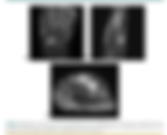
Synovial hemangioma of the wrist with cystic invasion of trapezoid and capitate bones Zhao, X., Qi, C., Chen, J., Li, H., Zhang, Y., & Yu, T. Level of Evidence : 5 Follow recommendation : 👍 Type of study : Diagnostic/Therapeutic Incidence : Rare Topic : Synovial Haemangioma - Diagnosis and treatment This is the answer for the case study from last week. The patient was an 18 year old male who had been experiencing pain and swelling in the back of the wrist in the last 2 years. Objectively, there was a 3x3 cm non-pulsatile mass in the back of the wrist. Extension range of movement had a deficit of 20 degrees. X-ray was impeccable, however, computer tomography and MRI scans revealed an ill-defined soft tissue mass between scaphoid, trapezoid, and capitate. Following surgery, it was possible to make a diagnosis of wrist synovial haemangioma. Synovial haemangiomas are rare benign tumours which usually affect children or young adults. Only 300 cases have been reported in the literature, most of which occurred in the knee. Symptoms vary and intermittent pain may be present or absent. Disclaimer: This publication was reviewed and assessed by one reviewer only and it reflects their interpretation. Readers should come to their own conclusions by reading the original article. Clinical Take Home Message : Hand therapists should refer young children or teenagers for x-rays and ultrasound when there is evidence of an irregularly shaped, soft mass which appears to or is reported to have grown over time. The likelihood of identifying a synovial haemangioma is extremely rare, however, this work up would help differentiating among different conditions including ganglion cyst, rheumatoid arthritis, haematomas associated with haemophilia, infections or other rare forms of cancer. URL : https://www.jhandsurg.org/article/S0363-5023(18)30316-2/fulltext Available through The Journal of Hand Surgery (American Volume) for HTNZ members. Available through EBSCO Health Databases for PNZ members. Abstract Synovial hemangiomas (SHs) are rare lesions of the joints or tendon sheaths that are difficult to diagnose. We present the case of an 18-year-old man with an SH in the wrist joint. Physical examination revealed a slightly tender, ill-defined, nonpulsatile soft mass, 3 cm × 3 cm in size on the dorsal aspect of the left wrist. Computed tomography showed an irregular, ill-defined, soft tissue mass in the expanded joint space, which was formed by the scaphoid, trapezoid, and capitate bones. Magnetic resonance imaging showed the typical features of SH and also revealed cavitary erosion of the scaphoid, trapezoid, and capitate bones. An open arthrotomy was performed via a dorsal approach, and the mass was excised. The histological examination findings were consistent with the diagnosis of SH.